Source: McGovern Institute News
Small and tucked away under the cerebral hemispheres toward the back of the brain, the human cerebellum is still immediately obvious due to its distinct structure. From Galen’s second century anatomical description to Cajal’s systematic analysis of its projections, the cerebellum has long drawn the eyes of researchers studying the brain. Two parallel studies from MIT’s McGovern institute have recently converged to support an unexpectedly complex level of non-motor cerebellar organization, that would not have been predicted from known motor representation regions.
Historically the cerebellum has primarily been considered to impact motor control and coordination. Think of this view as the cerebellum being the chain on a bicycle, registering what is happening up front in the cortex, and relaying the information so that the back wheel moves at a coordinated pace. This simple view has been questioned as cerebellar circuits have been traced to the basal ganglia and to neocortical regions via the thalamus. This new view suggests the cerebellum is a hub in a complex network, with potentially higher and non-motor functions including cognition and reward-based learning.
A collaboration between the labs of John Gabrieli, Investigator at the McGovern Institute for Brain Research and Jeremy Schmahmann, of the Ataxia Unit at Massachusetts General Hospital and Harvard Medical School, has now used functional brain imaging to give new insight into the cerebellar organization of non-motor roles, including working memory, language, and, social and emotional processing. In a complementary paper, a collaboration between Sheeba Anteraper of MIT’s Martinos Imaging Center and Gagan Joshi of the Alan and Lorraine Bressler Clinical and Research Program at Massachusetts General Hospital, has found changes in connectivity that occur in the cerebellum in autism spectrum disorder (ASD).
A more complex map of the cerebellum
Published in NeuroImage, and featured on the cover, the first study was led by author Xavier Guell, a postdoc in the Gabrieli and Schmahmann labs. The authors used fMRI data from the Human Connectome Project to examine activity in different regions of the cerebellum during specific tasks and at rest. The tasks used extended beyond motor activity to functions recently linked to the cerebellum, including working memory, language, and social and emotional processing. As expected, the authors saw that two regions assigned by other methods to motor activity were clearly modulated during motor tasks.
“Neuroscientists in the 1940s and 1950s described a double representation of motor function in the cerebellum, meaning that two regions in each hemisphere of the cerebellum are engaged in motor control,” explains Guell. “That there are two areas of motor representation in the cerebellum remains one of the most well-established facts of cerebellar macroscale physiology.”
When it came to assigning non-motor tasks, to their surprise, the authors identified three representations that localized to different regions of the cerebellum, pointing to an unexpectedly complex level of organization.
Guell explains the implications further. “Our study supports the intriguing idea that while two parts of the cerebellum are simultaneously engaged in motor tasks, three other parts of the cerebellum are simultaneously engaged in non-motor tasks. Our predecessors coined the term “double motor representation,” and we may now have to add “triple non-motor representation” to the dictionary of cerebellar neuroscience.”
A serendipitous discussion
What happened next, over a discussion of data between Xavier Guell and Sheeba Arnold Anteraper of the McGovern Institute for Brain Research that culminated in a paper led by Anteraper, illustrates how independent strands can meet and reinforce to give a fuller scientific picture.
The findings by Guell and colleagues made the cover of NeuroImage.
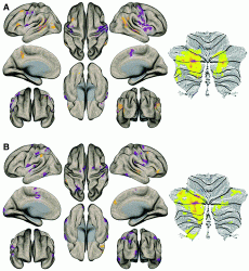
Anteraper and colleagues examined brain images from high-functioning ASD patients, and looked for statistically-significant patterns, letting the data speak rather than focusing on specific ‘candidate’ regions of the brain. To her surprise, networks related to language were highlighted, as well as the cerebellum, regions that had not been linked to ASD, and that seemed at first sight not to be relevant. Scientists interested in language processing, immediately pointed her to Guell.
“When I went to meet him,” says Anteraper, “I saw immediately that he had the same research paper that I’d been reading on his desk. As soon as I showed him my results, the data fell into place and made sense.”
After talking with Guell, they realized that the same non-motor cerebellar representations he had seen, were independently being highlighted by the ASD study.
“When we study brain function in neurological or psychiatric diseases we sometimes have a very clear notion of what parts of the brain we should study” explained Guell, ”We instead asked which parts of the brain have the most abnormal patterns of functional connectivity to other brain areas? This analysis gave us a simple, powerful result. Only the cerebellum survived our strict statistical thresholds.”
The authors found decreased connectivity within the cerebellum in the ASD group, but also decreased strength in connectivity between the cerebellum and the social, emotional and language processing regions in the cerebral cortex.
“Our analysis showed that regions of disrupted functional connectivity mapped to each of the three areas of non-motor representation in the cerebellum. It thus seems that the notion of two motor and three non-motor areas of representation in the cerebellum is not only important for understanding how the cerebellum works, but also important for understanding how the cerebellum becomes dysfunctional in neurology and psychiatry.”
Guell says that many questions remain to be answered. Are these abnormalities in the cerebellum reproducible in other datasets of patients diagnosed with ASD? Why is cerebellar function (and dysfunction) organized in a pattern of multiple representations? What is different between each of these representations, and what is their distinct contribution to diseases such as ASD? Future work is now aimed at unraveling these questions.